Street Address: Mater Misericordiae Hospital,
21-37 Fulham Road, PIMLICO Q 4812
Postal Address: P.O. Box 1417, Thuringowa Central, 4817
Phone: 07 4727 4097 Fax: 07 4727 4099
Pelvic organ prolapse
A pelvic organ prolapse occurs when there is a defect or weakness in the supporting structures or muscular structures of the pelvis. The pelvic floor muscle is a big hammock of muscles that supports the uterus, bladder and bowel. 1 in 9 women will develop a pelvic organ prolapse by the age of 80. There is a reported 29% recurrence rate of pelvic organ prolapse in women who have previous surgery for pelvic organ prolapse, with the surgical techniques of today this have been reduced.
Contributing causes of prolapses
Childbirth or pregnancy
Collagen deficiency (collagen is a natural protein that helps keep tissues plump and elastic)
Ageing and menopause
Being overweight
Chronic cough
Constant heavy lifting
Previous pelvic surgery
Chronic constipation
Complications
Symptoms:
Common complaints with a prolapse are
Sensation of "something coming down" when up and about, disappears when lying down.
Backache
Increased frequency of needing to void {which is probably due to incomplete emptying of the bladder.}
Problem emptying bowel
A feeling of fullness or pressure in the lower abdominal area, {which is probably due to venous congestion and pressure from the abdominal contents on a weak and inadequate pelvic floor}.
Sexual dysfunction and discomfort during intercourse.
Urinary stress incontinence.
While pelvic organ prolapses are not life threatening they can certainly have an effect on your overall wellbeing and quality of life.
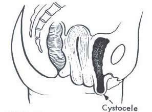
Cystocele or anterior wall prolapse
A cystocele is when there is a defect in the anterior wall supports and causes, the bladder to prolapse into the vaginal wall. This contributes to inability to empty the bladder properly and stress urinary incontinence.
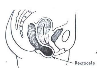
Rectocele or posterior vaginal wall prolapse
A rectocele is where the defect is in the posterior or back wall supports of the vagina and as a result the rectum bulges into the vagina. This can contribute to difficulty emptying the bowel as this causes a pocket to form and as a result faeces become lodged there, it is not uncommon for women with a rectocele to have to use perineal pressure to aid the emptying of their bowel. A rectocele can also cause a decreased stream when passing urine as the bulge presses up against the urethra (water pipe) thus obstructing the flow of urine.
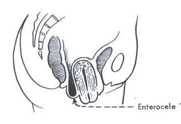
Enterocele
This is where the small bowel prolapses into the top of the vagina between the vagina and rectum, most commonly seen with a rectocele and or uterine prolapse.
Uterine prolapse
This is where the defect is in the structure that supports the womb, resulting in the cervix and uterus to prolapse into the vagina.
There are varying degrees of this prolapse
Grade 1: The uterus has dropped slightly and many women are unaware of the prolapse and are noticed during a routine pelvic check up.
Grade 2: The cervix and uterus has dropped further and can be felt or seen just inside the vagina.
Grade 3: The cervix and uterus can be seen or felt outside the vagina. This is the most severe of uterine prolapses and also knows as a procidentia.
Vaginal vault prolapse
The vaginal vault is the top of the vagina. With a vault prolapse this is where the vaginal wall loses its support from surrounding structure and the vagina falls in on it self. A vault prolapse can only occur to women who have had a previous hysterectomy. Vault prolapse statistics show increased risk of vault prolapse in women who had a hysterectomy for a uterine prolapse, the risk of this occurring are decreasing with new methods of surgery for prolapse repair.
There are varying degrees of prolapses and are graded as
Grade 1: prolapse is evident on examination but patient usually unaware of prolapse
Grade 2: Prolapse can be felt or seen at vaginal opening
Grade 3: Prolapse protrudes through the vaginal opening.
Urogynaecology Education prepared by: Prof. A. Rane & A. Corstiaans CNC: January 2004.